Intended for European healthcare professionals only
FGFR2 Helix

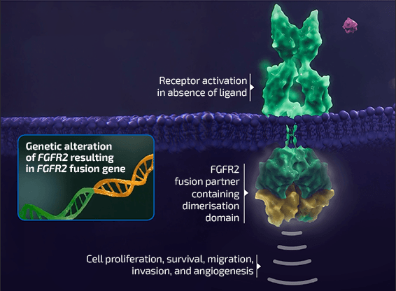
Fibroblast growth factor receptor 2 (FGFR2) fusions in intrahepatic cholangiocarcinoma (iCCA)

Genomic alterations in fibroblast growth factor receptors (FGFRs)

  • FGFRs are a family of receptor tyrosine kinases.1,2
    FGFR signalling pathways play a central role in multiple cellular processes, including cell proliferation, migration and survival1,2
  • Alterations in FGFR genes have emerged as tumourigenic drivers in cancers including iCCA, urothelial carcinoma, myeloid/lymphoid neoplasms and other malignancies1,3,4

  • FGFR amplifications, mutations and fusions have been observed in all FGFR subtypes (FGFR1–4).5 Chromosomal rearrangements involving FGFR2 – resulting in the creation of oncogenic fusion proteins – have frequently been identified in iCCA6
    • Gene fusions are a type of genomic alteration where two independent genes or portions of genes are juxtaposed, resulting in a hybrid gene7,8
    • The development of fusion proteins with oncogenic potential can result from gene fusion events involving a range of different partner genes7

FGFR genomic alterations

FGFR genomic alterations

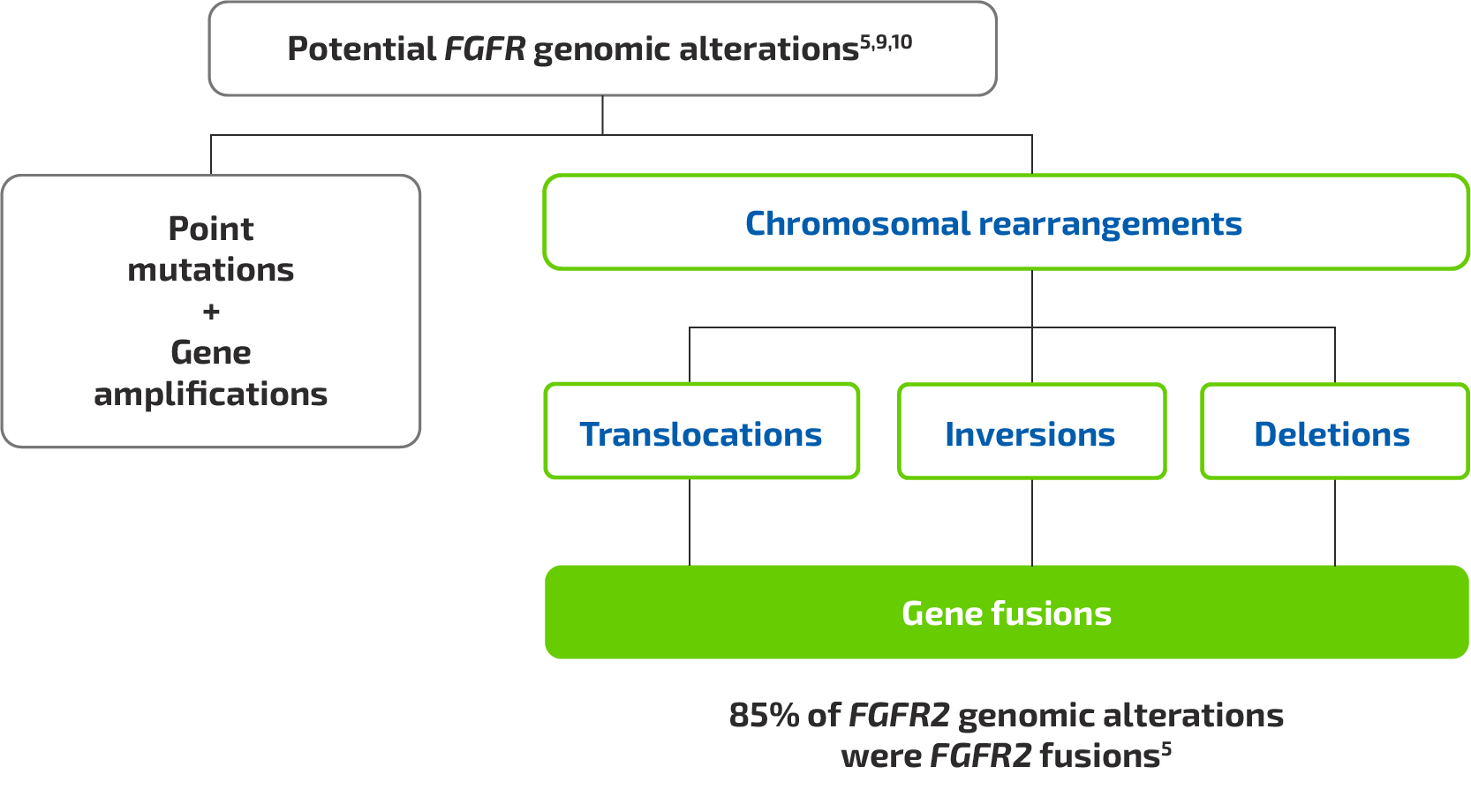
Figure based on Jain A, et al. 2018,5 Lowery MA, et al. 2018,9 and Shibata T, et al. 201810

FGFR2 fusions

  • FGFR2 fusions or rearrangements occur in 10–16% of iCCA cases5,11-13
  • FGFR2 fusions result in ligand-independent activation of downstream signalling pathways, leading to tumourigenesis1,14,15

Abnormal FGFR2 signalling pathway

Abnormal FGFR2 signalling pathway

Figure adapted from Babina IS, Turner NC. 2017,1 Moeini A, et al. 2015,14 and Touat M, et al. 201515

  • Tumour molecular profiling is necessary to identify FGFR2 fusions.5,9 Assessment for FGFR2 fusion positivity should be performed with an appropriate diagnostic test7
  • FGFR2 fusions involve a wide range of fusion partners.9
    To identify patients with FGFR2 fusion-positive cholangiocarcinoma (CCA), it is important to select an assay that:
    • Specifically detects FGFR2 fusions (distinct from FGFR2 point mutations)16,17
    • Detects FGFR2 fusions with a wide range of fusion partners16,17
  • The molecular diversity of CCA supports the use of DNA- or RNA-based next-generation sequencing (NGS) assays as standard to detect both known and novel FGFR2 fusions or rearrangements18

Testing methodologies for the detection of FGFR2 fusions

Click on a testing method to view the advantages and challenges

Least appropriate7,19–27
Most appropriate7,19–27
Least appropriate7,19–27
Most appropriate7,19–27

The European Society for Medical Oncology (ESMO) recommends routine use of NGS to detect FGFR2 fusions in advanced CCA28

Proposed algorithm of how FGFR2 fusion testing can be incorporated into a diagnostic work-up

Patient diagnosed with CCA Acquire patient tumour sample Oncologist to request FGFR2 fusion test Is in-house FGFR2 fusion testing available? No Pathologist to send sample to laboratory with FGFR2 fusion testing capabilities Yes Pathologist to perform FGFR2 fusion testing with an appropriate diagnostic test Pathologist to communicate FGFR2 fusion status to oncologist Oncologist to consider relevant treatment options for the patient

CCA, cholangiocarcinoma; FGFR2, fibroblast growth factor receptor 2

External quality assurance programmes are essential to ensure accurate and reliable clinical biomarker testing31

REFERENCES: 1. Babina IS, Turner NC. Nat Rev Cancer. 2017;17:318–32. 2. Turner N, Grose R. Nat Rev Cancer. 2010;10:116–29. 3. Pandith AA, et al. Urol Oncol. 2013;31:398–406. 4. Gallo LH, et al. Cytokine Growth Factor Rev. 2015;26:425–49. 5. Jain A, et al. JCO Precis Oncol. 2018;2:1–12. 6. Fangda L, et al. Cytokine Growth Factor Rev. 2020;52:56–67. 7. DeLuca A, et al. Int J Mol Sci. 2020;21:6856. 8. Latysheva S, Babu M. Nucleic Acids Research. 2016;10:4487–50. 9. Lowery MA, et al. Clin Cancer Res. 2018;24:4154–61. 10. Shibata T, et al. Cancer Sci. 2018;109:1282–91. 11. Ross JS, et al. Oncologist. 2014;19:235–42. 12. Farshidfar F, et al. Cell Rep. 2017;18:2780–94. 13. Graham RP, et al. Hum Pathol. 2014;45:1630–8. 14. Moeini A, et al. Clin Cancer Res. 2015;22:291–300. 15. Touat M, et al. Clin Cancer Res. 2015;21:2684–94. 16. Silverman IM, et al. Cancer Discov. 2021;11:326–39. 17. Barr FG. Expert Rev Mol Diagn. 2016;16:921–3. 18. Abou-Alfa GK, et al. Lancet Oncol. 2020;21:671–84. 19. Malka D, et al. EMJ Oncol. 2020;8:82–94. 20. Peter M, et al. Lab Invest. 2001;91:905–12. 21. Arai Y, et al. Hepatology. 2014;59:1427–34. 22. Abel H, et al. J Mol Diagn. 2014;16:405–17. 23. Beadling C. J Mol Diagn. 2016;18:165–75. 24. Hu L, et al. Biomark Res. 2014;2:3. 25. Maruki Y, et al. J Gastroenterol. 2021;56:250–60. 26. Serratì S, et al. Onco Targets Ther. 2016;9:7355–65. 27. Jennings LJ, et al. J Mol Diagn. 2017;19:341–65. 28. Vogel A, et al. Ann Oncol. 2023;34:127–40. 29. Patel T. Nat Rev Gastroenterol Hepatol. 2011;8:189–200. 30. Damodaran S, et al. Am Soc Clin Oncol Educ Book. 2015;e175–82. 31. Dufraing K, et al. Virchows Arch. 2021;478:553–65.