Intended for UK healthcare professionals only
Lungs

What is intrahepatic cholangiocarcinoma (iCCA)?

iCCA is a subset of cholangiocarcinoma (CCA)

CCA, the most common primary malignancy of the bile duct, is classified by anatomic origin into iCCA and extrahepatic CCA (eCCA). eCCA is further divided into 2 types:1-3

  • Perihilar CCA (pCCA) (also termed “Klatskin’s tumour”) is the most common form, accounting for 50–70% of all CCA cases1,3
  • Distal CCA (dCCA) accounts for 30–40% of total CCA cases1,3

iCCA originates in the bile ducts within the liver and accounts for <10% of CCA cases1,3

CCA is classified by anatomic origin3,4

Liver Image

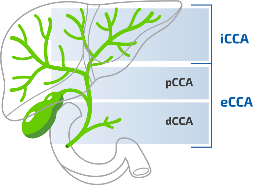
CCA, cholangiocarcinoma; dCCA, distal cholangiocarcinoma, eCCA, extrahepatic cholangiocarcinoma; iCCA, intrahepatic cholangiocarcinoma, pCCA, perihilar cholangiocarcinoma.

Figure adapted from Blechacz B. 2017.4

Incidence of iCCA

Throughout Europe, incidence rates of CCA range between 0.5/100,000 and 3.4/100,0006

Although rare, the incidence of iCCA is increasing worldwide7-9

The increasing incidence of iCCA may be due to factors including:7-9

  • Increasing burden of chronic liver disease
  • The potential role of environmental toxins
  • Improved diagnostic tools and imaging
  • Epidemiological trends such as increasing prevalence of diabetes, obesity and alcohol use

Worldwide incidence of CCA (cases per 100,000)6

Map of world showing incidence of CCA Map of world showing incidence of CCA

Data refer to the period 1971–2009. Where available, the more incident form (iCCA vs eCCA) and the temporal trend of incidence
(↑increasing trend; ↔ stable trend; ↓decreasing trend) have been reported.
CCA, cholangiocarcinoma; eCCA, extrahepatic cholangiocarcinoma; iCCA, intrahepatic cholangiocarcinoma.

Figure adapted from Banales JM, et al. 2016.6

Worldwide incidence of CCA (cases per 100,000)6

Austria

Austria

2.67 ↑eCCA>↑iCCA
(1990—2009)
Denmark

Denmark

1.27 ↓eCCA>↓iCCA
(1978—2002)
Finland

Finland

1.05
France

France

1.3 ↔eCCA>↔iCCA
(1976—2005)
Germany

Germany

Saarland 2 Hamburg 3 ↑eCCA=↑iCCA
(1970—2006)
Italy

Italy

3.36 ↑eCCA>↑iCCA
(1988—2005)
Poland

Poland

0.7
Spain

Spain

0.5
Switzerland

Switzerland

0.45
UK

UK

2.17 ↑iCCA>↓eCCA
(1971—2001)

Canada

Canada

0.35
USA

USA

1.6 ↑iCCA>↔eCCA
(2003—2009)

Costa Rica

Costa Rica

0.3
Puerto Rico

Puerto Rico

0.35

China

China

Shanghai 7.55 Qidong 7.45 iCCA>eCCA
Hong Kong 2.25 Guangzhou 0.97 iCCA>eCCA
Israel

Israel

0.3
Japan

Japan

Osaka 3.5 Hiroshima 3.05 ↓eCCA>↑iCCA
(1985—2005)
Philippines

Philippines

1.2 iCCA>eCCA
Singapore

Singapore

1.45 iCCA>eCCA
South Korea

South Korea

Gwangju 8.75 Daegu 7.25 Busan 7.1 ↑iCCA>↑eCCA
(1999—2005)
Taiwan

Taiwan

4.7 iCCA>eCCA
Thailand

Thailand

Northeast 85 North 14.6 Central 14.4 South 5.7 ↑iCCA>↔eCCA
(1998—2002)
Vietnam

Vietnam

0.1 iCCA>eCCA

Australia

Australia

0.43
New Zealand

New Zealand

0.4

Data refer to the period 1971–2009. Where available, the more incident form (iCCA vs eCCA) and the temporal trend of incidence
(↑increasing trend; ↔ stable trend; ↓decreasing trend) have been reported.
CCA, cholangiocarcinoma; eCCA, extrahepatic cholangiocarcinoma; iCCA, intrahepatic cholangiocarcinoma.

Unmet needs in iCCA

Patients with iCCA face significant clinical challenges, including a poor prognosis and limited treatment options10

In spite of improved technology, numerous diagnostic challenges persist in iCCA, including:10-12

As a result, 60–70% of patients are diagnosed with metastatic or unresectable disease, with a median survival of ~12 to 15 months.11 For the 30–40% eligible for resection—the only treatment with curative intent—the majority (~60–65%) experience disease recurrence11,13-15

Biopsy is necessary to confirm a specific diagnosis of iCCA12

As the scientific understanding of, and clinical management options for, iCCA evolve, it is critical to accurately diagnose iCCA and distinguish it from other cancers of the biliary tract; imaging alone is not sufficient12
REFERENCES: 1. Ghouri YA, et al. J Carcinog. 2015;14:1. 2. Ghidini M, et al. Cancer Manag Res. 2019;11:379–88. 3. Razumilava N, Gores GJ. Lancet. 2014;383:2168–79. 4. Blechacz B. Gut Liver. 2017;11:13–26. 5. Lowery MA, et al. Clin Cancer Res. 2018;24:4154–61. 6. Banales JM, et al. Nat Rev Gastroenterol Hepatol. 2016;13:261–80. 7. Khan SA, et al. Liver Int. 2019;39(suppl 1):19–31. 8. Kirstein MM, et al. Visc Med. 2016;32:395–400. 9. Valle JW, et al. Ann Oncol. 2016;27(suppl 5):v28–v37. 10. Blechacz B, et al. Nat Rev Gastroenterol Hepatol. 2011;8:512–22. 11. Bridgewater J, et al. J Hepatol. 2014;60:1268–89. 12. Banales JM, et al. Nat Rev Gastroenterol Hepatol. 2020;17:557–88. 13. Rizvi S, et al. Gastroenterology. 2013;145:1215–29. 14. Choi SB, et al. Ann Surg Oncol. 2009;16:3048–56. 15. Endo I, et al. Ann Surg. 2008;248:84–96.